Shape Of A Plant Cell
A institute cell consists of three distinct components:
(i) Cell wall
(ii) Protoplasm, and
(three) Vacuole.
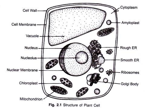
The protoplasm is the living function of the cell. Information technology is externally bounded by cell membrane or plasma membrane. The cytoplasm contains several cell organelles namely mitochondria, plastids, ribosomes, endoplasmic reticulum, lysosomes etc. (Fig. 2.1).
(i) Prison cell wa ll:
Cell wall is the non-living protective layer outside the plasma membrane in the plant cells, bacteria, fungi and algae. The synthesis of jail cell wall in controlled by Golgi bodies. In bacteria the cell wall is equanimous of protein and not-cellulosic carbohydrates while in virtually algae, fungi and all constitute cells, the cell-wall is formed of cellulose. Cell wall provides mechanical support and gives a definite shape to the cell. Information technology protects plasma membrane and helps in imbibition's of water and move of solutes towards protoplasm.
(ii) Protoplasm :
Protoplasm is the living, colourless, elastic, colloidal semi fluid substance nowadays in the cell. Protoplasm with non-living inclusions is called protoplast. Water is the chief constituent of an active protoplast and normally constitutes ninety% of the system. The remaining parts are organic and inorganic materials.
Each protoplast keeps itself in communication with neighbouring protoplasts through modest openings in the prison cell wall known as plasmodesmata. Protoplasm consists of cytoplasm and nucleus and is externally divisional by the jail cell membrane or plasmalemma.
(iii) Cell membrane :
Information technology is a thin film like pliable membrane, and serves as protective covering of the cell. Prison cell membrane mainly consists of proteins and lipids but in sure cases, polysachharides accept also been found. It facilitates the archway of nutrients into the cells and allows exit of nitrogenous wastes, regulates the passage of materials into and out of the cells. It controls and maintains differential distribution of ions inside and outside the cell.
(iv) Cytoplasm :
It is a jelly like fluid mass of protoplasm excluding the nucleus and surrounded by plasma membrane on the exterior. It is semi permeable in nature. The cytoplasm is composed of matrix; the membrane spring organelles and non-living inclusions like vacuoles and granules. The living cytoplasmic organelles are the site of various important metabolic activities such equally photosynthesis, respiration, poly peptide synthesis etc.
(v) Plastids :
Plastids are the largest cytoplasmic organelles bounded by double membranes.
There are iii types of plastids:
Leucoplast:
These are colourless plastids found in storage organs where lite is not available e.g. underground stem, roots and deeper tissues. Plastids without pigments are chosen leucoplasts. They shop starch, fats or proteins in meristematic, embryonic and germ cells.
Chromoplast:
Coloured plastids are chosen chromoplasts. These may contain ruby-red, yellowish, brown, purple, blue or dark-green pigments. These are by and large establish in petals of flowers and fruits.
Chloroplast:
In a plant cell, chloroplasts are the virtually prominent forms of plastids that contain chlorophyll, the green pigment. The chlorophyll enables the chloroplast to harness kinetic solar free energy and trap it in the form of potential energy. All living organisms direct or indirectly depend on them for energy. Chloroplast in enclosed in two smooth membranes separated past a distinct periplastidial space. The interior of chloroplast is differentiated into two parts— The Stroma and the Grana.
Stroma is the colourless ground substance that fills the chloroplast. It contains cholorophyll bearing double membranes lamellae that form flattened sac similar structures called thylakoides collectively chosen Grana. Quantasomes are the smallest units present on the inner surface of thylakoides capable of carrying out photochemical reactions (Fig. 2.2).

(half dozen) Ribosomes :
Ribosomes are the submicroscopic organelles. These are site of protein synthesis in the cell. These are found in all the cells either attached to the membranes of endoplasmic reticulum or scattered in the cytoplasm. The ribosomes are spheroidal bodies. In prokaryotic cells (Bacteria) the ribosomes are abour fifteen nm and in eukaryotic cells about 25 nm.
Ribosomes from prokaryotes exist as 70S units and ribosomes in eukaryotes exust as 80S units. A ribosome is formed of ii subunits – a big subunit and a pocket-size subunit. The modest subunit forms a sort of cap on the flat surface of large subunit. The 2 units of bacterial ribosome (70S) are represented by 50S and 30S subunits, and eukaryotic ribosomes (80S) are represented past 60S and 40S subunits.
The two subunits of ribosomes commonly exist complimentary in the cytoplasm and join only during protein synthesis when a number of ribosomes get fastened to mRNA in a linear mode. These groups or clusters of ribosomes are known equally Polyribosomes. The larger subunits (i.eastward. 60S and 50S) are attached to the membrane of endoplasmic reticulum and the smaller subunits are then bound to larger subunits. The cleft separating the two subunits lies parallel to the membrane. The messenger RNA is held by the smaller subunit, while tRNA molecule is jump to the larger subunit.
(vii) Mitochondria :
Mitochondria are sausage- shaped spherical or thread like organelles nowadays in the cytoplasm. They break downwardly the complex carbohydrates and sugars into usable forms and supply energy for the cell, they are also chosen as the powerhouse of the cell.
The mitochondria are surrounded by a double walled membrane known every bit outer and inner membranes. The spaces betwixt these two membranes are known equally perimitochondrial space. The outer membrane is smooth but the inner membrane is variously folded into sparse cristae. Inner membrane is covered with special particles called Oxysomes, these are the sites of aerobic respiration. (Fig. 2.ii).

(viii) Nucleus:
The nucleus is the nigh important role of the jail cell which regulates all metabolic and hereditary activities inside the cell It is more or less spherical, lying in the cytoplasm and occupying about two-thirds of the jail cell space. A typical nucleus is composed of the following structures (Fig. 2.2).
Nuclear membrane:
Information technology is a selectively permeable envelope-like construction surrounding the nucleolus and nucleoplasm. It is formed of two layers separated by a fluid-filled perinuclear space. The nuclear membrane disappears during prophase stage of nuclear division and reorganizes during telophase. Information technology regulates the passage of ions, small molecules and macromolecules of ribosomal subunits, mRNA, t RNA etc.
Nucleoplasm:
Nucleoplasm is the transparent semifluid basis substance formed of a mixture of proteins, phosphorus and some nucleic acids. Chromatin fibres or the chromonemata remain suspended in the nucleoplasm.
Chromatin network:
Chromatin fibres course a network in the nucleoplasm called chromatin net piece of work or nuclear reticulum. Chromatin fibres are the sites of primary genetic material which controls all the activities of the cell, metabolism and heredity. During cell division, the chromatin threads condense and form thick chromosomes.
Nucleolus:
The nucleolus is a spherical body lying in the nucleoplasm closely associated to the nucleolar organizer region of the chromosome. It was beginning described by Schleiden in 1838. It contains large amount of RNA though DNA is besides present. Its main function is the synthesis of ribosomal RNA (rRNA), which helps in synthesis of ribosomes.
(ix) Golgi Body :
Golgi torso is also referred to as golgi complex or golgi appliance. It plays a major office in transporting chemic substances in and out of the prison cell. It has three singled-out components flattened sac or cisternae, clusters of transition tubules and vesicles and large vesicles or vacuoles. Golgi is mainly associated with secretory activity of the cell. Information technology is also associated with the concentration, storage, condensation and packaging of materials for export from the cell across plasmalemma.
(x) Endoplasmic Reticulum :
Endoplasmic reticulum (ER) is the connecting link betwixt the nucleus and cytoplasm of the plant cell. Basically, it is a network of interconnected and convoluted sacs that are located in the cytoplasm. Based on the presence or absence of ribosomes, ER can be of shine or rough types. The sometime type lacks ribosomes, while the latter is covered with ribosomes. Overall, endoplasmic reticulum serves as a manufacturing, storing and transporting structure for glycogen, proteins, steroids and other compounds.
(xi) Lysosomes :
Lysosomes are tiny membrane-bound, vesicular construction of cytoplasm which enclose hydrolytic enzymes and perform intracellular digestion. These are also known as suicidal bags. These are constitute in all animal cells but only in few plant cells.
Types of Lysosomes :
Based on function or stage of digestion lysosomes are of post-obit four types:
(i) Main lysosomes are newly formed lysosomes with hydrolytic enzymes.
(ii) Secondary lysosomes are newly formed by the fusion of phagosomes and primary lysosomes. Here contents of phagosome are digested or hydrolysed.
(iii) Remainder bodies are exhausted secondary lysosomes. These contain undigested remains.
(iv) Autophagic vacuoles are formed by the fusion of chief lysosomes with jail cell organelles from prison cell's own cytoplasm. This brings almost autodigestion of cell or its organelles.
Functions :
Lysosomes bring almost digestion of extracellular and intracellular materials. Because of this basic character lysosomes perform following functions:
(i) Lysosomes of granular leucocytes or macrophages devour foreign substances and microbes which enter the cell and guard our torso against infection.
(ii) Lysosomes remove worn out cell organelles, dead cell and provide energy during starvation by controlled breakdown of stored food substances.
(iii) During dedifferentiation of tissues and cells, the lysosomes deliquesce the specialized parts of the cells. This helps in regeneration of damaged tissue or damaged part and germination of bone from cartilage.
(iv) The lytic enzymes of sperm acrosome aid in the penetration of sperm into the ovum.
(v) During metamorphosis, reabsorption of diverse larval structures like external gills of tadpole, the tail of polliwog in frog or the larval organs in the pupa of diverse insects is brought about by autolytic activity of lysosomes.
(vi) Lysosomes bring about cellular breakdown associated with ageing.
(7) Lysosomes may crusade cancer by breaking down chromosomes.
( xii ) Peroxisomes:
Peroxisomes occur widely both in establish and brute cells. They are spherical or ovoid bodies surrounded past a single membrane. It contains certain oxidative enzymes, used for the metabolic breakdown of fatty acids into elementary sugar forms. In light-green plants, peroxisomes aid in undergoing photorespiration.
( xiii ) Vacuoles:
Vacuoles are sap- filled vesicles in the cytoplasm. These are surrounded by a membrane called tonoplast. In a plant cell, in that location tin can be more than i vacuole; nevertheless, the centrally located vacuole is larger than others.
Tonoplast is a semi permeable membrane; it enables the vacuoles to concentrate and store nutrients and waste products. It facilitates the rapid commutation of solutes aid gases betwixt the cytoplasm and adjoining fluids.
(fourteen) Cilia and Flagella :
Cilia and flagella are motile hair -similar appendages on the complimentary surfaces of the cells. These are cytoplasmic processes and create water currents, nutrient currents, act as sensory organs and perform several other functions of the cell. Cilia and flagella tin be differentiated on the footing of their size, withal, other physiological and morphological characteristics are nigh the aforementioned.
The main difference between cilia and flagella are as follows:
Cilia and flagella are cylindrical processes projecting from the free surface of the cell. These originate from their basal bodies embedded in the cytoplasm. The basal bodies course their kinetic centres. A ciliam or flagellum consists of a longitudinal axoneme enclosed in a spiral sheath of cytoplasm and a plasma membrane continuous with the jail cell membrane.
Shape Of A Plant Cell,
Source: https://www.biologydiscussion.com/plants/structure-of-plant-cell-explained-with-diagram/2511
Posted by: christoffersothemnioncy64.blogspot.com


0 Response to "Shape Of A Plant Cell"
Post a Comment